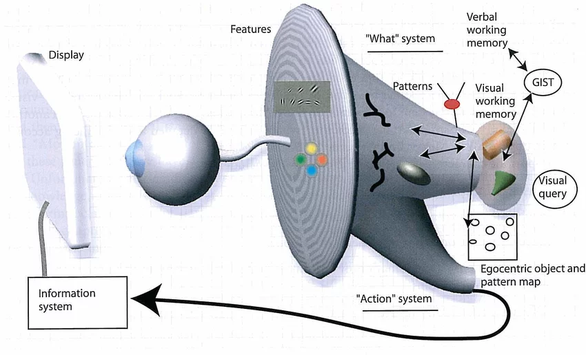
The notion of visual information processing is an effort to understand the complex integration of an organism’s internal functioning and its apparent behavior. Many researchers have laid the concept of the interrelation of the brain’s neuronal activity and our conscious visual experience. Whereas the inaugural researchers in the field of cognitive psychology have made efforts to outline the anatomy and physiology of the human brain’s visual areas’ response to external stimuli, many recent researchers have started to imply more on decoding the neural code of visual perception and the concept of hierarchies of visual computations. This paper will focus on the processing of visual information in the occipital cortex, two medical conditions that affect visual information processing and the latest trends that have aided in a better understanding of this phenomenon.
The human brain is a complex matter that has designated its several regions for interpretations of specific stimuli, which may overlap with each other sometimes. In regard to the visionary stimulus, initially, it sits on the retina and axons exiting the retina that makes up the optic nerve take this information to the visual cortex i.e. the occipital lobe of the brain. Two relays for visual information are the thalamus (the primary relay) and the superior colliculus (which helps in the movement of the gaze). The occipital lobe, which is not only responsible for perceiving but also for analyzing the external stimulus, is divided into many hierarchal sub-regions, each contributing to the processing of the simple to complex visual inputs in a descending manner. These areas decipher the ‘What’ and ‘Where’ information from the external stimulus. The top area of the ‘What’ region tells us the type of external stimuli like a face or any object in particular. The top area of the ‘Where’ region integrates the saccadic eye movement that helps us with the placement of our gaze on any object.
Visual information processing skills can be divided into many subareas. They include visual-spatial skills, which is the ability to judge the environment in context to oneself, visual analysis skills, which is the ability to detect, recollect and manipulate information from the already stored visual memory, and visual motor skills, also known as the ‘hand-eye coordination, is the synergistic movement of the hands in accordance to the visual stimulus. Many medical conditions can affect the visual processing of the brain, disrupting it at any level between the eyes and the brain. Two of these conditions are visual processing disorder or dyslexia and age-related macular degeneration affecting figure-ground discrimination.
Dyslexia is the disorder of the brain’s area called the lateral and ventral geniculate nuclei of the thalamus, which makes it hard for the person to read, write and spell. This condition has varying degrees of difficulties ranging from the inability to interpret and analyze moving visual stimuli to and differentiation of simple phonetics. Visual information is relayed at the lateral geniculate nucleus of the thalamus, specifically the Magnocellular and the Parvocellular cells. In dyslexics, either these cells do not recognize their division of labour or overlap in function, therefore leading to difficulties in visual motion detection. Neuropsychotherapy, phonetic instructions, auditory stimulus and proper nutrition have shown positive results in coping with dyslexia throughout life.
Age-related macular degeneration (AMD) can lead to disruption in figure-ground discrimination of the adult. It results in irreversible degenerative changes in the central part of the retina called the ‘macula’, leading to changes in perception of visual stimulus. Figure-ground discrimination is the ability to differentiate multiple objects in relation to the foreground and background stimulus, and as age progresses, their ability to discriminate the object from their fore/background decreases. Research have shown that white space around the objects can help in improving the contrast between different objects hence aiding in the geriatric population’s visual rehabilitation.
Many researchers in the current time have yielded new ways to study the concept of visual information processing. Similarly, Sterzer et al. present a critical review of the Continuous Flash Suppressing (CFS) technique and present knowledge about the science of how the interocular suppressed information is processed in the brain and the controversies relating to it. When two highly contrasting images competing for dominance are presented to the eyes, one of the images is suppressed in one eye unconsciously for a few minutes. This phenomenon helps in understanding the visual processing of either social, emotional or materialistic stimuli when there is no conscious awareness. On the other hand, this technique had its drawbacks at the same time, such as the disappearing image in the binocular rivalry was documented to reappear on and off in between, leading to false positive findings or the suppression of the stimulus was too deep, negating the stimulus completely resulting in false negative results(Aboudib, Gripon, & Coppin, 2016).
Future studies should avoid subjective documentation of the suppressing image, and the examiner should hold the threshold of the stimulus suppression hence altering it accordingly. Another technique that has emerged from the (CFS) is the “breaking-CFS”, which measures the time for how much the suppressed stimulus remains in the state of unawareness. Although this technique is still debatable, neuroimaging can help solve this by conjoining the neural responses to the initially suppressed stimulus and the time is taken by the stimulus to reappear. (Sterzer, Stein, Ludwig, Rothkirch, & Hesselmann, 2014)
In recent times, many researchers, under the light of visual information processing, are aiming to combine the science of the ventral stream in order to achieve a better understanding of machine learning algorithms for artificial intelligence. This research paper focuses on providing a skeletal framework for visual information acquisition by providing two prodigies in order to achieve the aforementioned goal, and that is cortical magnification and selective visual attention. The former helps in administering the saccadic eye movements for stable gaze movements, while the latter being the primary part of the visual system, helps in decreasing the amount of visual stimulus entering the eye. This framework can be administered flawlessly without spatial disruption and the difficulty of adjusting the size of the image.
Moreover, it can help in understanding the interrelation of the human eye’s distance to an object for a given visual task and decreases the amount of visual input received by the brain. In the coming times, this research paper offers to be used as the base for object recognition processors that are attention-based; also, other commonly used architectures processing visions can use this framework in a more efficient way.
